
ECG Wave and Screen Examples (Figure_13)
|
Procedure |
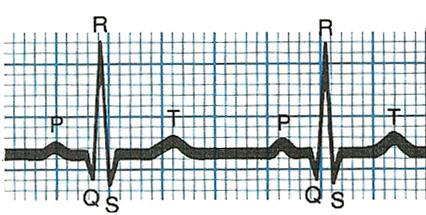 Example of Normal Wave (labeled) The normal wave is labeled with different parts p, q, r, s, and t. The "r" is the primary part used for beat detection. The "t" can cause beat detection problems if too "tall". |
Example of Baseline "Wander" This is an indication that the lead is moving relative to the patient. Try replacing the electrode and placing it over a more stable site (i.e. over bone). For Stress and Holter, taping the lead wires down and making a Stress loop and taping it down can help significantly. |
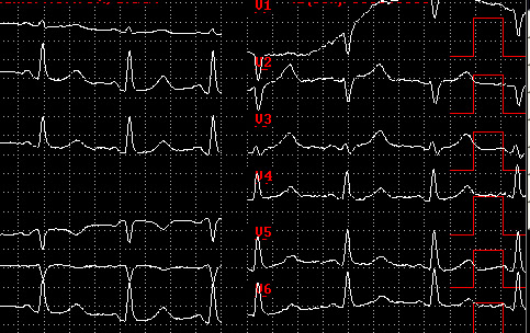
Example of V2 Lead Disconnect This is an indication that the lead wire or electrode are not making good contact. The electrode should be replaced. |
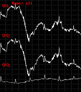
Example of "Noise" in 1st and 2nd Channel This is an indication that the lead wires or electrodes are not making good contact. The electrodes should be replaced. Note that an electrode site common to both channels may be the actual problem. |
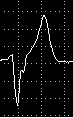
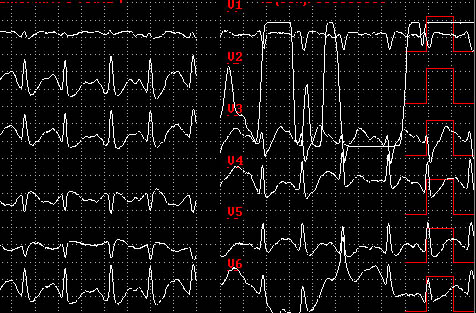
Example of "VE Beat" Ventricular events (VE, PVC) are wider than normal ECGs. The wave can have many forms. This example illustrates a biphasic wave with the leading part going negative. Others can have positive leading parts. |

Example of a "VE Beat" Couplet (2 in a row) |
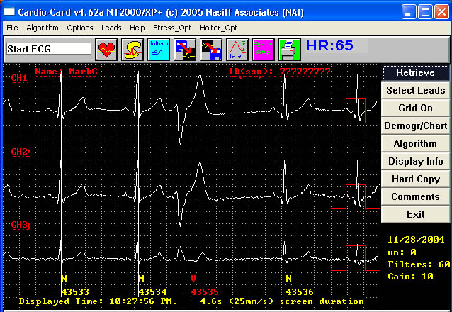
Example of Holter "Marks" and "Beat Codes" Note the vertical line goes through the "R" waves. At the bottom of the beat are the "Beat Codes" and "Beat Numbers". If you have marks on the "T" waves or a significant number of "R" waves are not marked, go to the CardioDatabase - Holter Monitoring Special Considerations and Instructions (Section III) of the Holter Monitor Index. |